This post is also available in:
![]()
![]()
![]()
![]()
Iceberg Diseases
Factsheet name: Investigation of thin ewes to determine if poor body condition is due to an iceberg disease
Need / Issue: There is a need for information on iceberg diseases in relation to poor ewe body condition
Introduction:
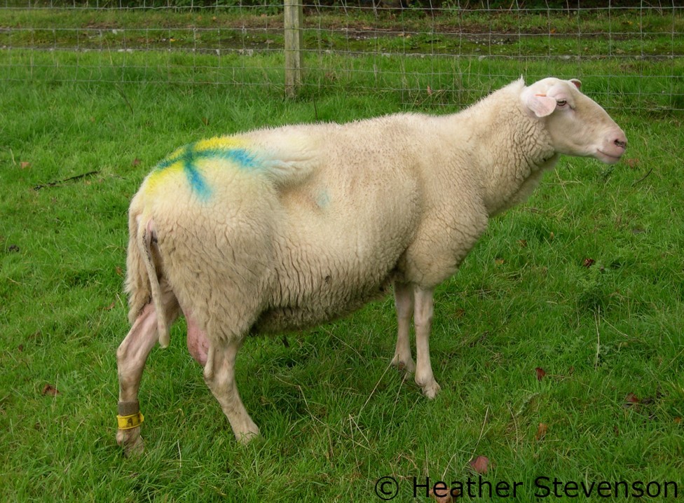
The iceberg diseases that cause poor ewe body condition are Johnes disease, maedi visna (MV) and ovine pulmonary adenocarcinoma (OPA/Jaagsiekte). In all cases the incubation period following infection is long. Usually only a small number of adult sheep in a flock show clinical signs. They may remain bright and continue to eat but lose weight without an obvious explanation. Thin ewes are often presented for investigation if they fail to gain weight within one to two months of weaning despite good nutrition. Thin ewes that are scanned not in lamb are also good candidates for investigation. Testing can include postmortem examination of typical cases, and this may be the most cost-effective way to reach a diagnosis. Blood and faecal samples can be collected from live ewes.
Johnes Disease
Johnes disease is caused by the bacteria Mycobacterium avium paratuberculosis. It is passed in faeces and can survive in the environment for weeks to months. Weight loss in a ewe that remains bright and continues to eat is often the only clinical sign of Johnes disease. In contrast to the disease in cattle diarrhoea is rarely seen. Possible routes of investigation include serology, faecal tests (ZN smear or PCR) +/- postmortem examination +/- histopathology. Lambs are at greatest risk of infection and factors that can reduce disease challenge include:
- Cull known positive ewes.
- Don’t keep replacements from known positive ewes.
- Reduce faecal contamination of fleece and teats around lambing (bedding, dagging).
- Cull thin ewes pre lambing.
- Tight lambing period so that weaning is not delayed.
- Beware of the risk from cattle colostrum.
- Move out of the lambing shed/field as soon as possible.
- Reduce stocking density from lambing to weaning.
- Provide mains water from clean troughs.
- Wean as early as possible onto low(er) risk grazing (not grazed by adult sheep for as long as possible).
- Beware of the potential risk from cattle.
- Consider the direction of water courses and run off.
- Avoid spreading slurry on grazing land.
- Ensure good nutrition and parasite control.
- Don’t overgraze – the Johnes bacteria can survive in soil for 1 year.
- Split flock by age and retain replacements from the younger group.
- Draft/cull ewes a year early.
- Vaccination – will not eradicate disease but will reduce losses and environmental challenge.
Not all measures will be practical or appropriate in all flocks.
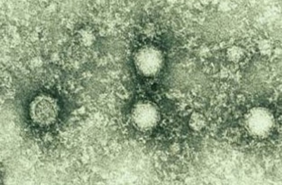
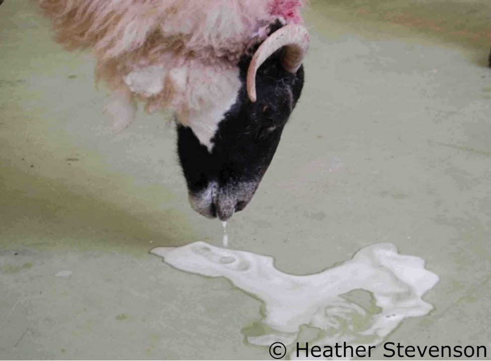
Maedi Visna
Maedi/Visna is a slow progressive viral disease that spreads mainly vertically (through the ingestion of colostrum from infected ewes) or horizontally (through respiratory secretions) in flocks. It is caused by strains of the genus Lentivirus. Housing and high stocking densities increases the possibility of airborne transmission through respiratory droplets during exhalation, sneezing, and coughing. The virus affects several organs leading to clinical disease with signs from:
- the respiratory system (dyspnea and increased respiratory rate in adult sheep),
- the udder (”hard bag” type of mastitis with decreased milk production mainly the first days postpartum), and/or
- the joints (arthritis with “swollen” joints, less common in sheep),
- the neurological system (ataxia, paresis, weakness in hind limbs, incoordination or, in heavier cases, total paralysis).
Since there is no treatment or vaccine, the only way to control the disease is to apply specific management strategies on farm level. The early diagnosis of infected animals is crucial for the control of the disease. The diagnosis is based on the detection of antibodies against the viral proteins. However, since antibodies usually found in animals older than 2 years, PCR-based diagnostic protocols have been proposed that detect the virus itself or its precursors stage, before the antibodies appearance. Annual, biannual, or more frequent blood sampling from the breeding stocks and serological and molecular testing are needed for the diagnosis of the infected animals.
Two disease control strategies are proposed depending on the flock prevalence:
- Infection rate less than 10% – “examination and slaughter” of positive animals and their offspring. Specific blood tests are available.
- Infection rate greater than 10% – immediate removal of new-borns from their mothers, administration of colostrum from healthy animals (or from cows or commercially available immunoglobulin preparations or commercial colostrum for lambs) and application of artificial feeding.
Embryo transfer from infected ewes could be used before culling. It should be emphasized that for the success of the program it is important to keep the replacement animals in a separate place and to avoid their contact with the adult animals of the rest flock.
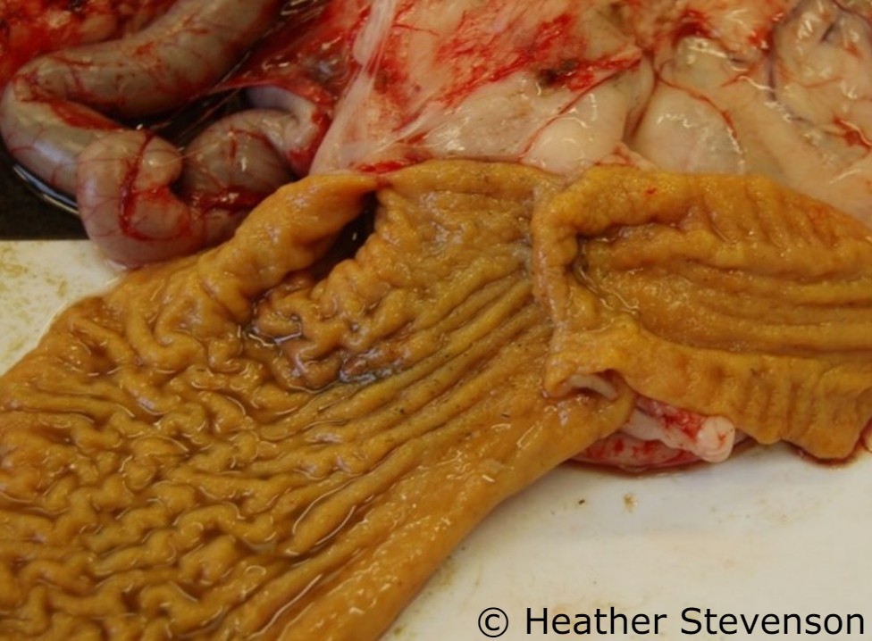
Ovine Pulmonary Adenocarcinoma
OPA is a viral disease that causes lung tumours often with the accumulation of excess fluid in the airways. Infection is spread in the nasal discharges of infected sheep. Housing, high stocking densities and trough feeding can increase the rate of spread. Weight loss may be the only clinical sign but evidence of respiratory disease such as exercise intolerance, increased respiratory rate, abdominal lift and open-mouthed breathing may be seen. There may be a nasal discharge or an occasional soft cough. Affected sheep are often treated with antibiotics for suspected pneumonia but respond poorly to treatment. OPA predisposes sheep to Pasteurella pneumonia, and as a result they may be found dead while still in good body condition. An accurate blood test for OPA is not available. A PCR test has been developed and can be used on nasal swabs but is expensive with poor sensitivity. Ultrasound scanning of lungs can be used but won’t detect the smallest tumours. If used as the basis of a test and cull programme scanning should be repeated after 3 to 6 months.
It is therefore important to investigate cases of sudden death and poor body condition with no obvious cause. Other ways to reduce the spread of OPA can include:
- Don’t retain lambs from known positive ewes. They may be infected via nasal discharges from the ewe or via milk/colostrum.
- Minimise periods of housing/trough feeding.
- Keep younger tups in a separate group.
- Good boundary biosecurity.
- Buy from trusted sources.
Topic: health
Production: Dairy / Meat
Animal Category: Ewes
This post is also available in:
![]()
![]()
![]()
![]()
